Tag: difference between light & electron microscope
Difference Between Light Microscope And Electron Microscope
Difference Between Light Microscope And Electron Microscope: Microscopes are indispensable tools in the world of science and research, enabling us to explore the microscopic world and uncover intricate details of specimens.
Two primary types of microscopes used for this purpose are the light microscope (LM) and the electron microscope (EM).
Despite their common goal of magnifying objects, these microscopes differ significantly in their principles of operation, capabilities, and the types of specimens they can examine. Here, we highlight the key differences between light microscopes and electron microscopes.

Difference Between Light Microscope And Electron Microscope
1. Principle of Operation:
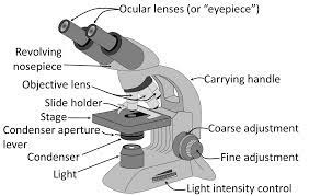
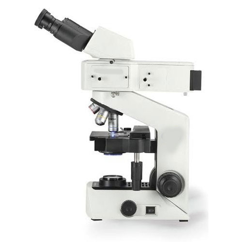
- Light Microscope (LM): LMs use visible light to illuminate and magnify specimens. They employ glass lenses to bend and focus light, revealing the specimen’s details. LMs are also known as optical or compound microscopes.
- Electron Microscope (EM): EMs use a beam of electrons instead of light to magnify specimens. Electrons have much shorter wavelengths than visible light, allowing for much higher magnification and resolution.
2. Magnification:
- Light Microscope (LM): LMs typically offer magnifications of up to around 1,000 times the specimen’s actual size. They have limitations in achieving higher magnification due to the wavelength of visible light.
- Electron Microscope (EM): EMs can achieve magnifications exceeding 50,000 times, and some advanced EMs can go beyond 2,000,000 times. This exceptional magnification is due to the short wavelength of electrons.
3. Resolution:
- Light Microscope (LM): LMs have limited resolution, typically around 200 nanometers (nm). This means that two points closer than 200 nm will appear as a single point.
- Electron Microscope (EM): EMs offer much higher resolution, often reaching below 0.1 nm. This allows them to reveal fine details of specimens at the atomic and molecular level.
4. Specimen Type:
- Light Microscope (LM): LMs are suitable for viewing living and non-living specimens such as cells, tissues, bacteria, and larger structures. They can observe specimens in their natural, hydrated state.
- Electron Microscope (EM): EMs are used to examine non-living specimens, as the process requires a vacuum. They are ideal for studying ultra-thin sections of cells, viruses, nanomaterials, and inorganic structures.
5. Sample Preparation:
- Light Microscope (LM): Sample preparation for LMs is relatively simple. Specimens are typically mounted on glass slides, often stained to enhance contrast.
- Electron Microscope (EM): EMs require more complex sample preparation, including fixation, dehydration, embedding in resin, and ultra-thin sectioning. Samples must withstand the vacuum and electron beam.
6. Color:
- Light Microscope (LM): LMs provide color images as they use visible light. Stains and dyes can enhance contrast and highlight specific structures.
- Electron Microscope (EM): EMs produce grayscale images since they use electrons. Different shades of gray represent variations in electron density within the specimen.
7. Cost and Accessibility:
- Light Microscope (LM): LMs are generally more affordable and widely accessible in educational and research settings.
- Electron Microscope (EM): EMs are complex and expensive instruments, often found in specialized research institutions and laboratories.


In summary, light microscopes are versatile tools suitable for studying a wide range of biological and non-biological specimens in their natural state, whereas electron microscopes provide unparalleled resolution and are indispensable for detailed examinations of nanoscale structures but require more extensive sample preparation and are primarily used for non-living specimens. The choice between the two depends on the specific research objectives and the nature of the specimens being studied.
Read More
- Molar Mass Of Aluminium
- Molecular Mass Of Glucose
- Latent Heat Of Water
- Difference Between Work And Power
- Difference Between Gravitation And Gravity
Frequently Asked Questions (FAQs) Difference Between Light Microscope And Electron Microscope
1. What is the main difference between a light microscope and an electron microscope?
The primary difference is in the type of radiation used for imaging. Light microscopes use visible light, while electron microscopes use a beam of electrons.
2. How do light microscopes and electron microscopes differ in terms of magnification
Light microscopes typically offer magnifications of up to around 1,000 times the specimen’s actual size, while electron microscopes can achieve magnifications exceeding 50,000 times and even up to 2,000,000 times.
3. What is the resolution difference between these microscopes?
Light microscopes have limited resolution, typically around 200 nanometers (nm), whereas electron microscopes offer much higher resolution, often below 0.1 nm.
4. Can light microscopes be used for studying living specimens?
Yes, light microscopes are suitable for observing living specimens such as cells, tissues, and bacteria in their natural, hydrated state.
5. Are electron microscopes suitable for studying living specimens?
No, electron microscopes require a vacuum environment, making them unsuitable for living specimens. They are primarily used for non-living specimens.